
Biofilms
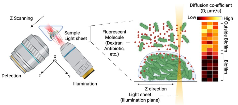
Biofilms are dense communities of bacteria held together by a self-produced matrix, and that matrix makes them much harder to treat than free-swimming bacteria. Z-scanning light-sheet imaging FCS lets us measure how fluorescent molecules such as dextrans, antibiotics and DNA nanoparticles diffuse in and around live biofilms.
The resulting diffusion maps show molecules slowing sharply as they enter the biofilm, revealing where and how strongly its structure limits penetration.
People
In collaboration with Mark Schembri, The University of Queensland, Australia.
Bacterial screening assay
There is a growing need for new antimicrobials in personal hygiene products. Compounds that attack bacterial membranes are attractive because bacteria cannot easily change their membrane composition, but existing screening methods are slow.
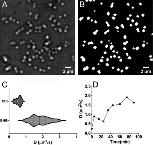
We combined imaging total internal reflection FCS (ITIR-FCS) with a segmentation algorithm that automatically finds bacterial cells, and measure how compounds change membrane dynamics in live bacteria across concentrations and incubation times. The assay reports membrane activity in under 30 minutes and produces dose–response curves within 2 hours, detecting activity at lower concentrations and an order of magnitude faster than commonly used susceptibility tests.
People
In collaboration with Procter & Gamble, Singapore.
Recent publications
- The biofilm matrix of uropathogenic Escherichia coli contributes to antimicrobial resistance by reducing molecular transportKulkarni R, Lian ZJ, Nhu NTK, Blaskovich MAT, Wohland T, Schembri MAnpj Biofilms and Microbiomes (2026). doi.org/10.1038/s41522-026-01174-6Biofilms
- Decoding interactions between biofilms and DNA nanoparticlesSousa A, Kulkarni R, Johannessen M, Wohland T, Skalko-Basnet N, Obuobi SBiofilm (2025). doi.org/10.1016/j.bioflm.2025.100260Biofilms
- A live bacterial screening assay for membrane-active antimicrobial compounds using imaging fluorescence correlation spectroscopyZhu S, Wang Y, Chong SMS, Wohland TAnalytical Chemistry (2025). doi.org/10.1021/acs.analchem.4c05698Screening assay