

How the cytoskeleton organises the membrane
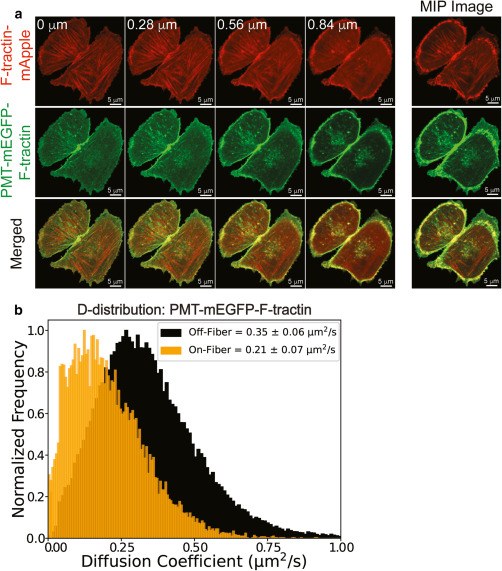
The plasma membrane is not a uniform fluid: the actin cytoskeleton beneath it pins and partitions membrane molecules. Combining imaging FCS with dual-colour imaging, we follow a membrane-anchored probe that binds actin and compare its mobility on and off actin fibres.
The probe diffuses markedly slower on fibres (about 0.21 µm²/s) than between them (about 0.35 µm²/s), showing how cytoskeletal pinning shapes the nanoscale organisation of the cell membrane.

Epidermal growth factor receptor (EGFR)
EGFR is a receptor tyrosine kinase that controls cell growth and development, and its misregulation is implicated in many cancers. How the receptor is organised before activation, as monomers, preformed dimers or larger oligomers, is still debated.
Using single-wavelength FCCS and imaging FCS we showed that EGFR forms preformed dimers in resting cells and that these complexes depend on cell line, temperature and membrane location. We are now using number and brightness analysis, drug treatments and EGFR mutants to quantify higher-order oligomers and the role of the membrane in regulating the receptor.
Read more on the full EGFR page.
Membrane-active peptides
Antimicrobial and amyloid-forming peptides act by binding to and disrupting lipid membranes. We use imaging FCS to follow how these peptides change membrane dynamics in model and live cell membranes.
Our recent work showed that the phenylalanine residues of the antimicrobial peptide aurein 1.2 act together to govern how it disrupts membranes. For our earlier work on peptides such as hIAPP, see peptide-membrane interactions.
Recent publications
- Ghosts on the membrane: cytoskeletal pinning influences nanoscale cell membrane organizationPandey S, Wohland TBiomolecules (2026). doi.org/10.3390/biom16040596Cytoskeleton and membrane
- Synergistic effect of phenylalanine residues governs the mechanism of membrane disrupting action of aurein 1.2Saadatmand ZN, Zhu S, Mechler A, Wohland TJournal of Medicinal Chemistry (2026). doi.org/10.1021/acs.jmedchem.5c02612Membrane-active peptides