
Biophysical Fluorescence Laboratory
Deciphering dynamics at different biological length and time scales by fluorescence
Research
Hover a topic for a short summary. Click for projects, people and papers.
Cells and cell membrane
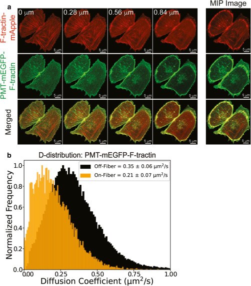
Imaging FCS combined with dual-colour imaging lets us see how actin fibres shape membrane dynamics in live cells. A membrane-anchored probe that binds actin diffuses markedly slower on fibres than between them, revealing how the cytoskeleton organises the plasma membrane.
Explore cell projects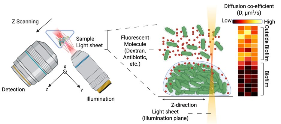
Bacteria and biofilms
Z-scanning light-sheet imaging FCS maps how fluorescent molecules such as dextrans and antibiotics diffuse in and around bacterial biofilms. Diffusion coefficient maps show molecules slowing sharply once they enter the biofilm, revealing how its structure limits penetration.
Explore bacteria projects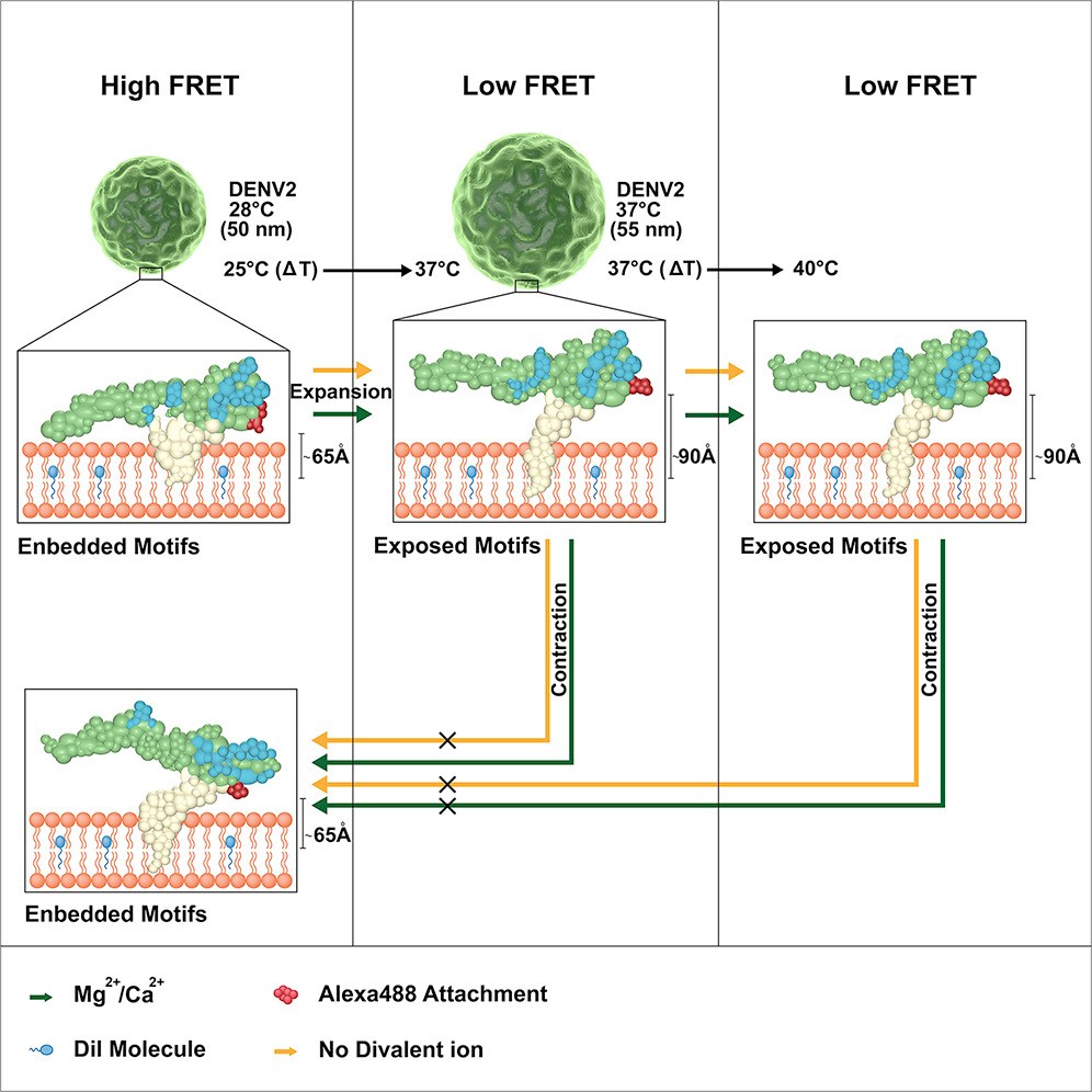
Viruses
We observe hitherto unseen conformational changes and structural dynamics of the DENV2 envelope that are influenced by both temperature and divalent cations. Our results show that for DENV2 and DENV1 the intrinsic dynamics, but not the specific morphologies, are correlated with viral infectivity.
Explore virus projects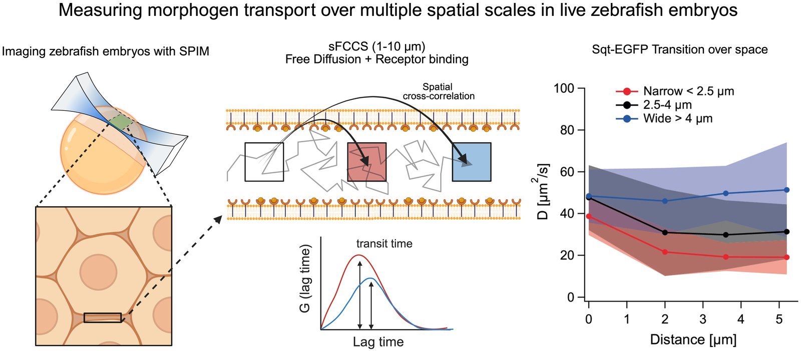
Zebrafish
Using SPIM-based spatial fluorescence cross-correlation spectroscopy over 1 to 10 µm, we follow how the morphogen Squint moves through the extracellular space of live zebrafish embryos. Separating free diffusion from receptor binding shows that transport slows with distance in narrow gaps between cells but stays fast in wider spaces.
Explore zebrafish projects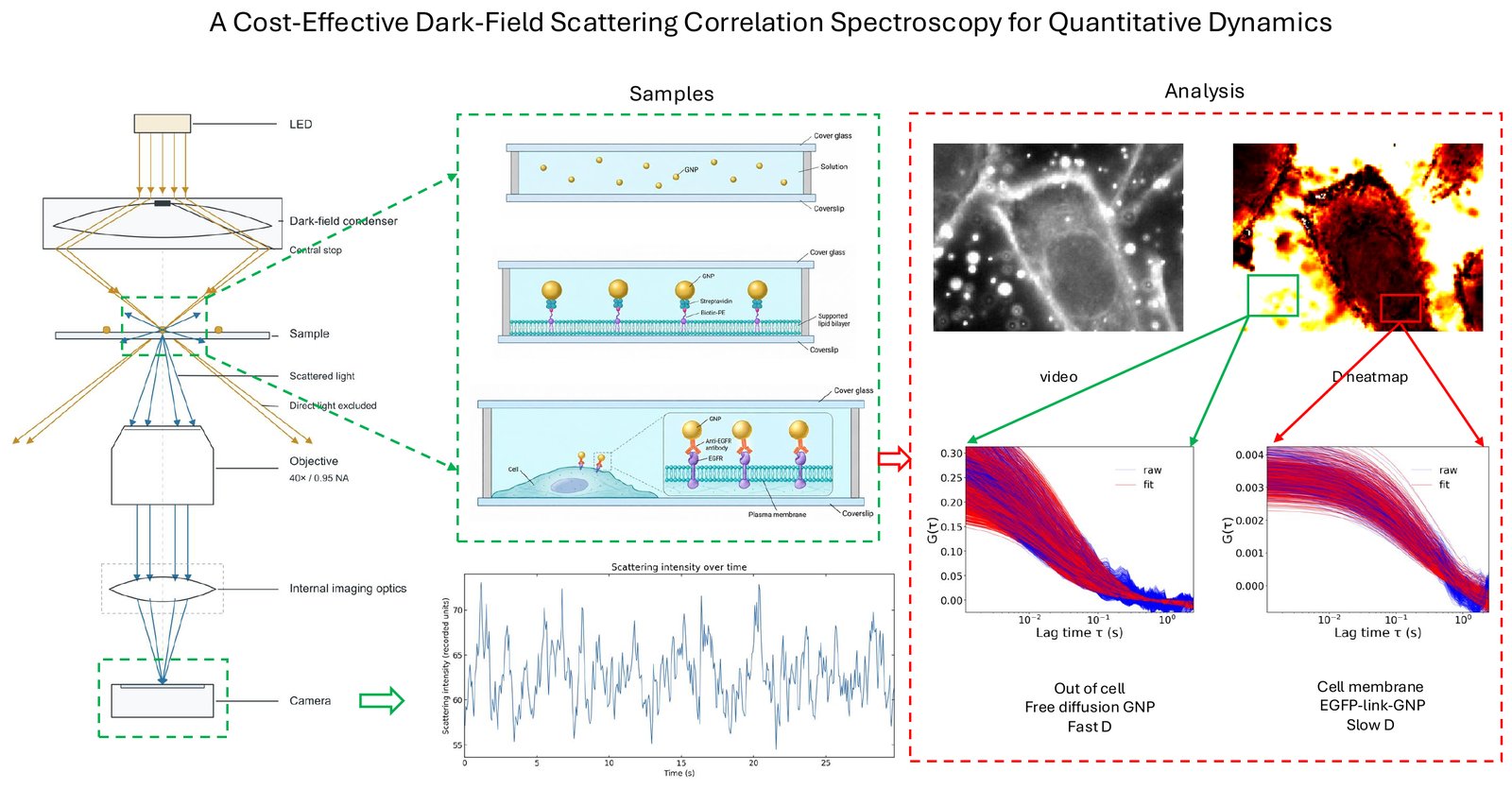
Instrumentation
We developed a cost-effective dark-field scattering correlation spectroscopy platform for quantitative measurement of nanoparticle dynamics. Validated across GNPs in solution, supported lipid bilayers, and cell membranes, the platform enables detection of mobility changes and provides a foundation for antimicrobial screening based on membrane disruption.
Explore our instruments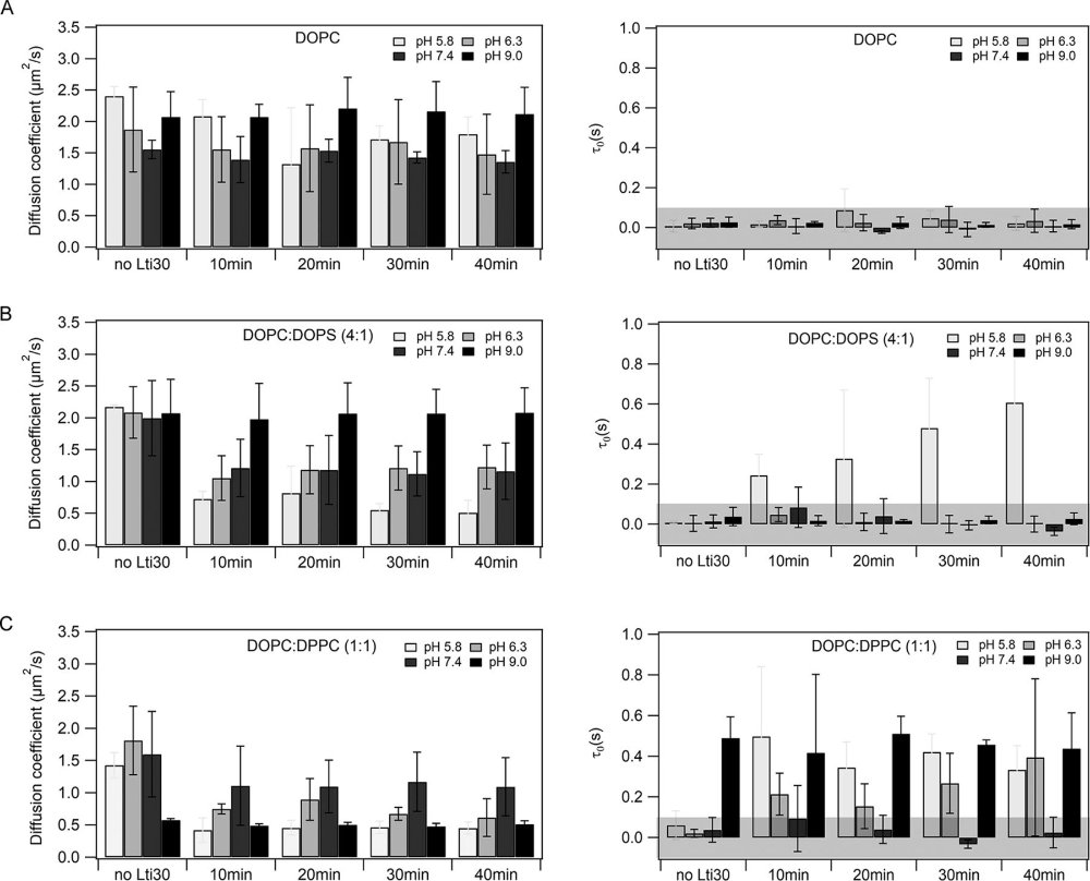
Bilayers
Using imaging total internal reflection FCS and molecular dynamics simulations on supported lipid bilayers, we showed how the plant dehydrin Lti30 protects membranes during cold and drought stress: it binds lipids electrostatically in a pH-dependent way, cross-linking them and restricting their mobility.
Explore bilayer projects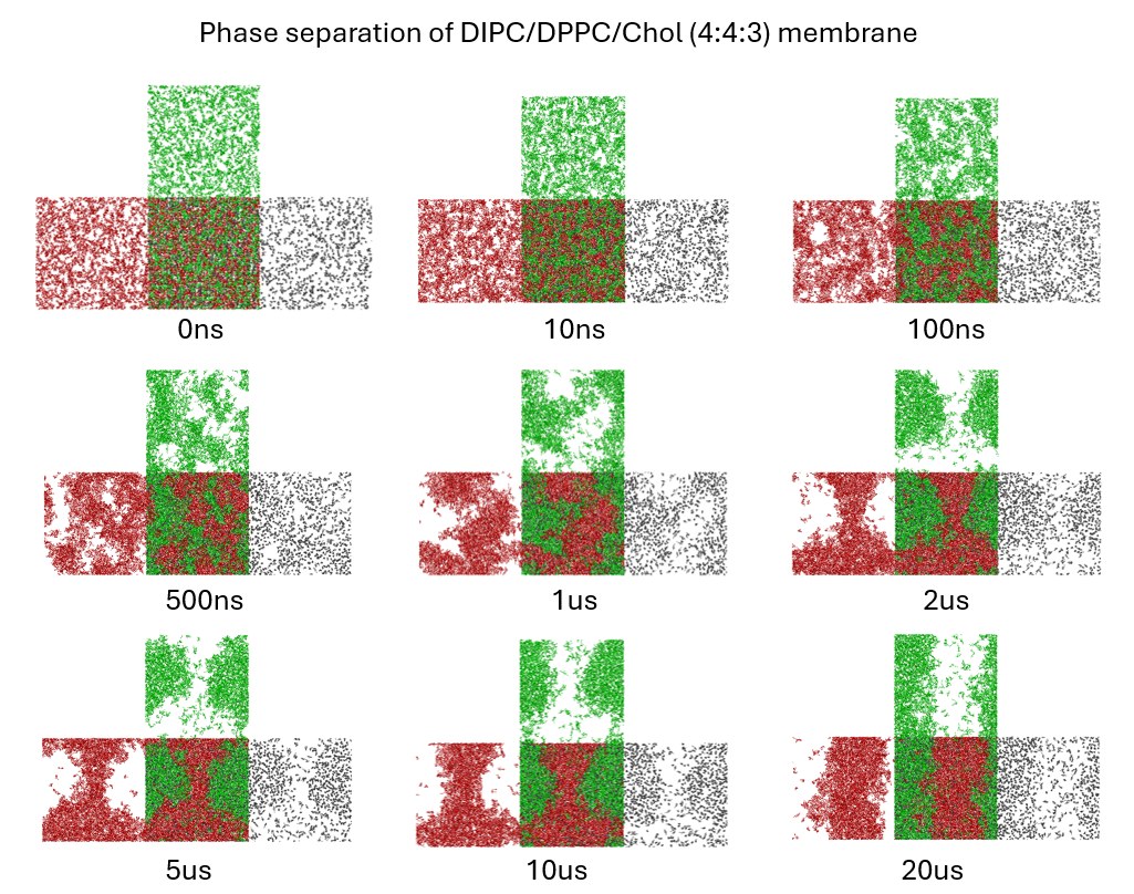
Simulations
Martini 3 coarse-grained MD snapshots (0 ns to 20 µs) of a DIPC/DPPC/Chol (4:4:3) bilayer undergoing spontaneous phase separation.
AI and ML
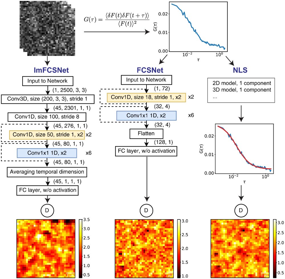
The deep-learning networks FCSNet and ImFCSNet predict diffusion coefficients more precisely than conventional fitting while needing far less data, cutting a typical Imaging FCS measurement from about 100 MB to 5 MB and opening the way to real-time measurements.
Explore AI and ML projectsResources
Free software and tutorials for anyone working with FCS.
Software
External tools and tutorials