

30 June 2026 – winner of CBIS microscopy image competition
Tirtha Das Banerjee
(PI: Professor Antonia Monteiro, NUS Department of Biological Sciences)
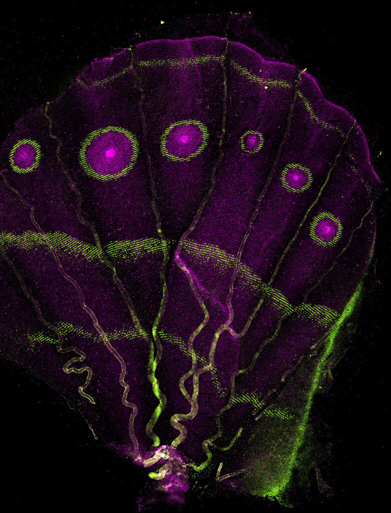
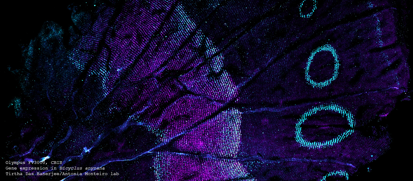
Genes encoding transcription factors Optix (green) and Spalt (magenta) light up the eyespot rings of a Bicyclus anynana pupal wing. These two genes define eyespot colors today by reusing an ancient gene network that patterns insect wing veins, dating back ~400 million years. Evolution’s favorite trick: reuse, not reinvention. Old genes, new beauty.
Imaged with Olympus FV3000, CBIS Light Microscopy Core
30 June 2024 – winner of CBIS microscopy image competition
Zhang Shiyu
(PI: Assoc Prof Liou Yih-Cherng, NUS Department of Biological Sciences)
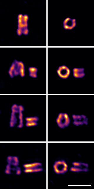
Tracking the newborn centriole in synchronized HeLa cells: HeLa cells were synchronized at G1/S and then released. Centriole growth was monitored every 240 minutes for 12 hours until the cells reached G2/M. Ultra-expanded and stained for acetylated α-tubulin (mpl-Inferno), both longitudinal (left 4 panels) and orthogonal (right 4 panels) views reveal the dynamic lengthening of newborn centrioles. Row 1: 0 minutes post-release. Row 2: 240 minutes post-release. Row 3: 480 minutes post-release. Row 4: 720 minutes post-release. Scale bar, 0.5 μm
Imaged with ZEISS LSM 900 with Airyscan 2, CBIS Light Microscopy Core
April 2024 – CBIS microscopy image competition
Submission deadline 31 May 2024 · terms and conditions
August 2023 – New in-situ cryotomography technique discerns nanometer-scale structures in chromatin with broad applications in cell biology.
Principal investigator: Lu Gan | lab website | read the paper
September 2022 – By knowing the physics of electron-matter interactions, a single 2D transmission electron micrograph can now be used to reconstruct 3D structures at nanometer resolution.
Principal investigator: Duane Loh | lab website | read the paper
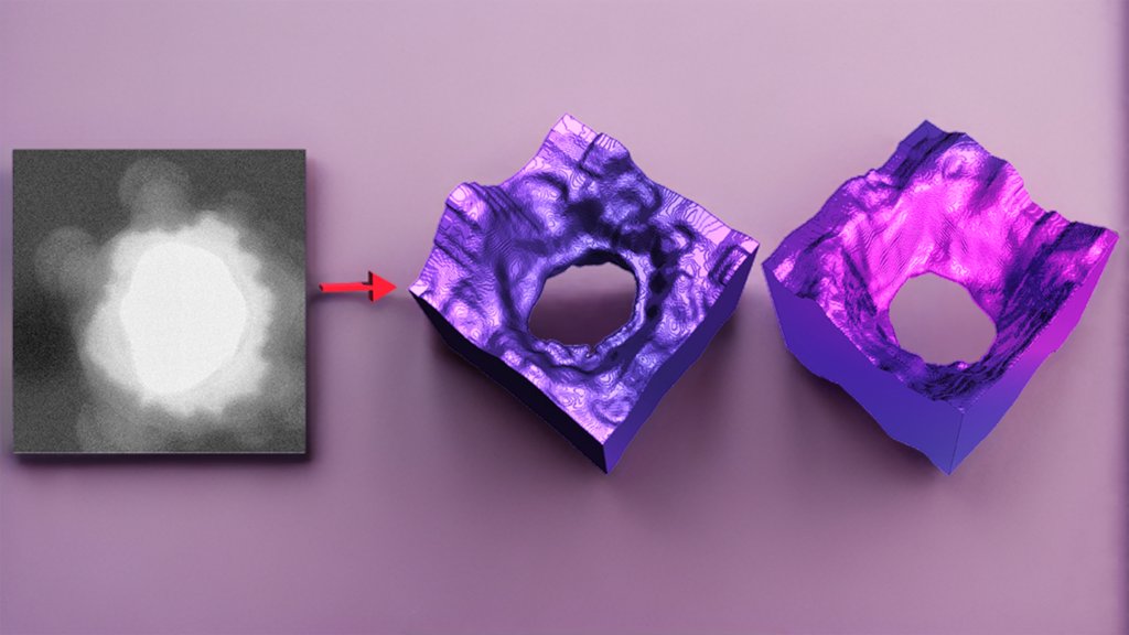
©Deepan Balakrishnan