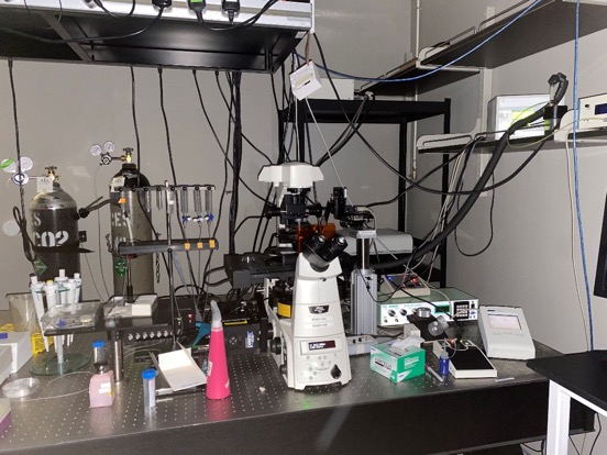
CBIS LM Core: Widefield Fluorescence/Dark Field/TIRF
The Plant Journal
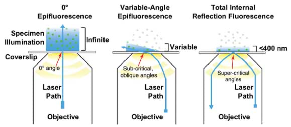
In total internal reflection fluorescence (TIRF), the laser contacts the coverslip at a very shallow angle and gets completely reflected by the glass surface adjacent to the sample before it can hit the sample (right). This generates in the sample a very shallow evanescent field of the same wavelength as the laser, which excites the fluorophores in the sample.
One can also steepen the laser’s angle of incidence so it passes through the coverslip into the sample at a very shallow angle (centre). This essentially gives widefield fluorescence with much less out-of-focus illumination, yielding clearer images with less photodamage.
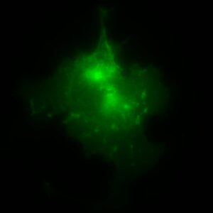
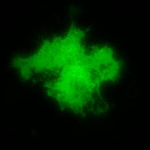
Mammalian cells with GFP localization in widefield (upper) and TIRF (lower). Sample preparation: Divya Dhasmana; principal investigator: Thorsten Wohland, Biophysical Fluorescence Laboratory; imaging: Tong Yan, Light Microscopy Core, Centre for Bioimaging Sciences.
TIRF has a large field of applications, such as single molecule imaging, imaging secretion processes, Fluorescence Recovery After Photobleaching (FRAP, FLIP), photoactivation, etc.
Comparison of LM techniques:
|
Method |
Excitation |
Detection |
Optical Sectioning |
Application |
|
Widefield |
Whole volume |
Whole sample |
No |
Thin fluorescence sample |
|
Confocal |
Single beam cone illumination |
Focal plane |
350-500nm |
Optical sectioning for both thin and thick sample |
|
TIRF |
Evanescent field |
Bottom plane adjacent to coverslip |
100-200nm |
Membrane dynamics |
Help
Widefield fluorescence user guide
Darkfield user guide
Features
- Nikon Eclipse Ti inverted microscope, with Perfect Focus System (PFS), Nikon TI-S-EJOY
- Transmitted light for DIC, phase contrast and dark field
- Wide field:
- Ti-Filter blocks: Ti Filter DAPI, GFPHQ, TRITC
- light Source: XCite 200DC mercury
- Laser mode:
- (TIRF1) Diode module: 405nm 100mW, 488nm 150mW, 561nm 100mW, 640nm 100mW
- Dichroic mirror: Di01-R405/488/561/635
- Emission Filters: 435/40, 520/35, 609/54, 692/40
- Laser illuminator: iLas2 laser illuminator for ring TIRF and targeted laser action
|
Objective list |
Phase Contrast* |
DIC* |
|
Nikon PLAN FLUOR 10X/0.3 OFN25 PH1 DL |
Ph1 |
– |
|
Nikon PLAN APO 40X/0.95 λD |
– |
|
|
Nikon Apo TIRF 60X/1.49 0.13-0.21 DIC N2 (on request) |
– |
N2 |
|
Nikon Apo TIRF 100X/1.49 WD0.12, 0.13-0.20, DIC N2 (on request) |
– |
N2 |
|
*refer to corresponding condenser position |
||
- Detector: Hamamatsu Orca Flash 4.0 (liquid-cooled)
- Software: Metamorph
- Sample holders for 35mm petri dish, chamber slide, glass slide and multiple well plate