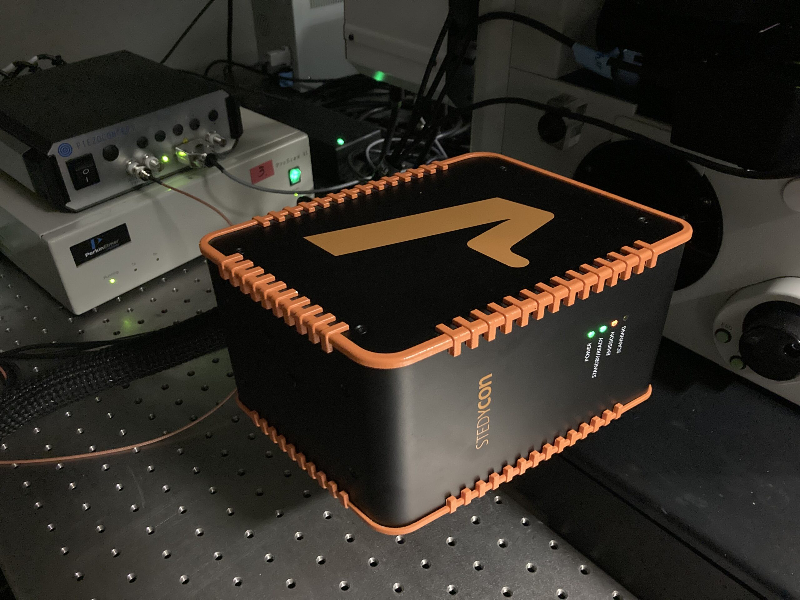
CBIS LM Core: Abberior STEDYCON
Stimulated emission depletion (STED) microscopy is a super-resolution technique that uses a doughnut-shaped laser beam of long wavelength to deplete the fluorescence signal to a tiny spot in the middle of the doughnut.
The higher the depletion laser power, the smaller the spot and the higher the resolution, but it causes more photodamage.
Images are super-resolution straight out of the microscope and do not require post-processing. However, deconvolution can be performed to further enhance resolution if desired.
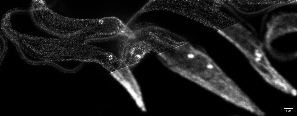 Trypanosoma brucei (STED versus confocal). Sample preparation: Assoc Prof Cynthia He, Principal Investigator, Molecular and Cellular Parasitology Lab, NUS; imaging: Tong Yan, CBIS Light Microscopy Core.